
Empirical discovery across biological, pharmaceutical, and material sciences is fundamentally bounded by optical resolution. From the identification of emergent viral pathogens and targeted oncological therapies to the sub-micron inspection of semiconductor lithography, scientific progress requires precise visualization of the microscopic domain. The optical microscope has transformed from a simple seventeenth-century dual-lens configuration into an automated, digital data-generation node.
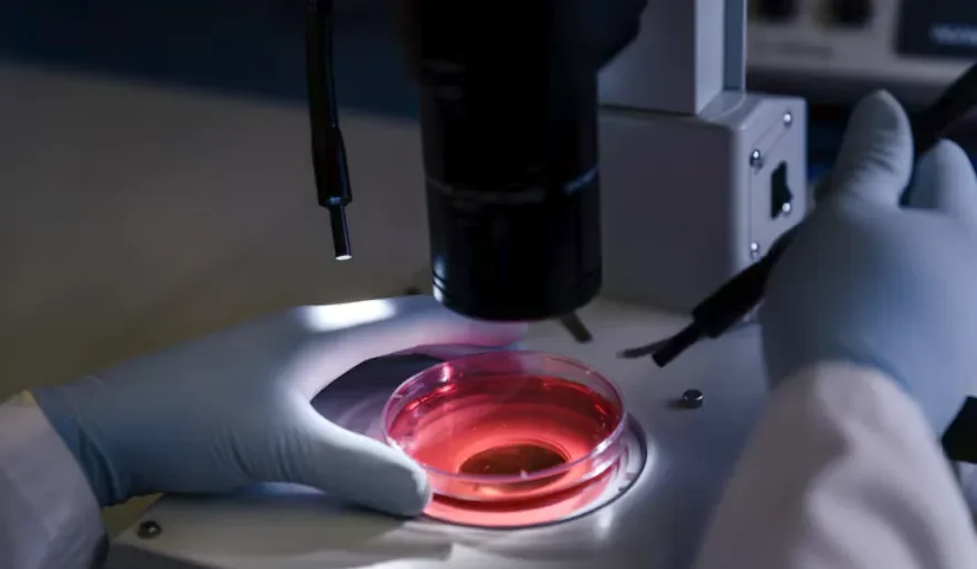
For clinical diagnosticians, university principal investigators, and industrial materials scientists, selecting and calibrating high-fidelity optical instrumentation is a critical prerequisite for data reproducibility and diagnostic validity. In high-throughput laboratories, the optical architecture of a facility dictates both the limits of resolution and the speed of scientific translation.
Comparative Matrix: Core Optical Microscopy Modalities
Different specimen types require distinct contrast mechanisms to overcome the physical limits of transparency, refractive index matching, and diffraction:
| Optical Modality | Contrast Mechanism | Specimen State | Primary Research Application |
| Brightfield | Differential light absorption through specimen | Fixed and chemically stained tissue sections | Routine clinical histology, biopsy evaluation, and hematology smears |
| Phase Contrast | Shifts in light wave phase converted to intensity variations | Unstained living cells in liquid culture | Real-time cell migration, mitotic monitoring, and stem cell maintenance |
| Differential Interference Contrast (DIC) | Nomarski prisms split polarized light into orthogonal shears | Unstained living cells or thick transparent sections | High-resolution optical sectioning of cell membranes, embryos, and living protozoa |
| Epifluorescence | Excitation of fluorophores releasing Stokes-shifted emission | Chemically labeled or genetically tagged specimens | Protein localization, antibody mapping, and targeted genomic FISH assays |
| Confocal Laser Scanning (CLSM) | Spatial pinhole aperture rejecting out-of-focus light | Thick intact tissues, embryos, and biomaterials | 3D volumetric reconstruction and multi-channel subcellular mapping |
The Physics of Resolution: Beyond Empty Magnification
A common misconception in microscopy procurement is prioritizing total magnification over resolving power. Magnifying an optically blurred image simply results in “empty magnification” with no added data. The definitive metric of optical performance is spatial resolution ($d$), governed by Ernst Abbe’s classical diffraction formulation:
$$d = \frac{\lambda}{2\,\text{NA}}$$
Where $\lambda$ represents the wavelength of the illuminating light, and $\text{NA}$ represents the Numerical Aperture of the objective lens:
$$\text{NA} = n \sin \theta$$
Here, $n$ is the refractive index of the imaging medium (air: $1.0$, water: $1.33$, immersion oil: $1.518$), and $\theta$ is the half-angle of the maximum cone of light collected by the objective. A high-NA objective gathers a wider angle of diffracted wave fronts, directly yielding a smaller value for $d$ and resolving closer adjacent structures.
Key Mechanical & Optical Metrics
-
Infinity-Corrected Optics: Modern research platforms utilize an infinity-corrected optical path between the objective and the tube lens. This design allows researchers to insert auxiliary optical blocks—such as fluorescence filter cubes, polarizers, and DIC prisms—without changing the mechanical tube length or introducing spherical aberrations.
-
Solid-State LED Illumination: Modern systems replace high-maintenance halogen and high-pressure mercury lamps with narrow-band, daylight-balanced Light-Emitting Diode (LED) sources. LEDs provide stable color temperatures across varying intensities, generate minimal radiant heat, and prevent thermal degradation during live-cell assays.
-
Ergonomic Mechanical Bases: Long microscopy sessions create ergonomic strain. High-end frames incorporate low-profile stage controls, adjustable torque mechanisms, and tilting trinocular viewing tubes to maintain neutral cervical alignment and reduce repetitive stress injuries.
The Digital Pathology Transition: Whole Slide Imaging and Computer Vision
The field of anatomical pathology is undergoing a shift from manual eyepiece observation to high-speed digital workflows:
[ Glass Slide Specimen ] ──► [ Automated High-NA Scanner ] ──► [ Gigapixel Image File ] ──► [ Cloud Telepathology & AI ]
 (Micro-biopsy mount)         (Continuous line scanning)        (SVS/NDPI format)          (Automated mitosis counts)
-
Whole Slide Imaging (WSI): Automated robotic stages and line-scan sensors digitize entire tissue sections at $20\times$ or $40\times$ optical equivalents within minutes. These gigapixel scans allow pathologists to zoom seamlessly from macroscopic tissue architecture down to sub-micron nuclear morphology.
-
Telepathology and Asynchronous Consultation: Digital slide archives enable instant remote consultations between clinical centers globally, eliminating transit delays for critical intraoperative frozen sections.
-
Machine Learning Assistance: Validated deep learning convolutional neural networks (CNNs) analyze digitized biopsy slides to identify regions of interest, quantify biomarker expression (such as HER2 or Ki-67), and segment rare circulating tumor cells, providing high-precision quantitative support for diagnostic teams.
Capital Allocation and Facility Integration
Selecting laboratory instrumentation requires evaluating the total lifecycle cost rather than initial hardware expenses alone:
-
Modular Upgradability: Optical stands should feature open mechanical architectures. A core facility can deploy a brightfield stand and later add epifluorescence carousels, motorized Z-drives, or environmental incubation chambers without replacing the microscope chassis.
-
Environmental Stability: High-resolution objectives are sensitive to floor-borne vibration and acoustic noise. Critical imaging suites require optical isolation tables and regulated HVAC systems to eliminate mechanical drift during extended time-lapse captures.
-
Software Portability: Image acquisition suites must support open microscopy environments (such as Micro-Manager or Bio-Formats), ensuring imaging metadata remains compliant with FAIR (Findable, Accessible, Interoperable, and Reusable) data standards.
Maintenance Standards: Preserving Optical Integrity
Preserving the resolving power of a research microscope requires strict operational hygiene:
-
Daily Köhler Alignment: Technicians must align the condenser height and center the field and aperture diaphragms before every imaging session. Proper Köhler illumination ensures uniform field brightness, eliminates internal reflections, and optimizes the balance between contrast and resolving power.
-
Oil Immersion Protocols: High-index immersion oil must be removed immediately after each session using dry optical lens tissue followed by high-purity isopropyl alcohol or specialized lens-cleaning solutions. Residual oil can harden over time, attracting abrasive dust and degrading the optical cement sealing the front lens elements.
-
Environmental Decontamination: Optical components in shared live-cell environments must undergo routine inspections for fungal spores and moisture condensation, which can permanently damage anti-reflective fluorite coatings.
Modern optical instrumentation serves as a vital bridge between physical specimens and quantitative, digital insights. By combining high-NA optics, multi-modal contrast systems, and high-speed digital detection, research institutions establish the reproducible optical foundation required to drive the next generation of scientific discovery.